Electrical Impedance Tomography — Lung Imaging During Parabolic Flight.
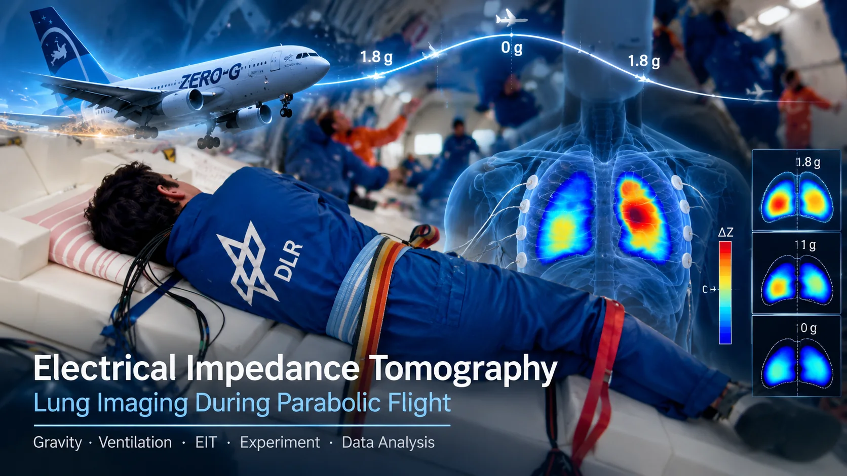
What happens to the distribution of air inside the human lungs when the apparent gravitational load changes? A parabolic aircraft makes it possible to investigate this question within seconds, moving from approximately normal gravity through hypergravity to short intervals of near-weightlessness. Electrical impedance tomography (EIT) offers a way to observe regional changes in lung aeration and ventilation without using ionizing radiation.
THE IDEA — MAKING AIR DISTRIBUTION VISIBLE
The lung is an electrically heterogeneous organ. Air is a poor electrical conductor, while blood and other tissues provide more conductive pathways. During breathing, changes in regional air content modify the electrical properties measured around the chest. EIT exploits these changes to obtain time-resolved images of the cross-sectional distribution of impedance-related quantities.
The experimental question is not simply whether a person can breathe in microgravity. It is how the distribution of ventilation and aeration responds when the gravity-related pressure gradients and mechanical loading of the lung change. Parabolic flight allows measurements under several loading conditions during one experimental session.
THE PHYSICS — CURRENTS, POTENTIALS AND CONDUCTIVITY
Electrodes arranged around the thorax inject small alternating currents and measure the resulting boundary voltages. Within a quasi-static approximation, conservation of charge and Ohm's law give
∇ · (σ(r) ∇φ(r)) = 0,
where φ denotes electric potential and σ electrical conductivity. For frequency-dependent tissue properties, the conductivity can be generalized to a complex admittivity, γ(r,ω) = σ(r,ω) + iωε(r,ω). In practice, electrode contact and the geometry of the thorax also influence the measured voltages.
Changing the current-injection and voltage-measurement electrode pairs provides many boundary measurements. Unlike computed tomography based on X-rays, EIT does not directly measure individual straight-line paths through the body. The current spreads throughout the conductive volume, so reconstruction requires a physical model of the electrical field.
FROM SIGNALS TO IMAGES — DATA ANALYSIS
Functional EIT usually reconstructs changes relative to a reference state. For sufficiently small changes, the forward model can be linearized:
ΔV ≈ J Δσ,
where ΔV is the vector of measured voltage changes, Δσ represents conductivity changes in the reconstruction domain, and J is the sensitivity (Jacobian) matrix. Recovering the conductivity changes is an inverse problem: measurements are incomplete and noise-sensitive, so regularization and assumptions about the geometry are needed.
During respiration, regional impedance changes provide a surrogate for regional ventilation. Repeated images can be evaluated over the breathing cycle, compared between lung regions, or grouped according to the aircraft's acceleration phase. However, reconstructed color patterns are not direct photographs of air, and their numerical interpretation depends on reconstruction method, reference, electrode configuration and motion artifacts.
Functional versus absolute EIT. Functional EIT emphasizes relative changes, such as tidal ventilation. Absolute EIT attempts to estimate the underlying conductivity or resistivity distribution itself and is more sensitive to calibration and modeling assumptions. Both approaches appear in the Göttingen group's research literature [1–3].
THE EXPERIMENT — LUNG MEASUREMENTS IN PARABOLIC FLIGHT
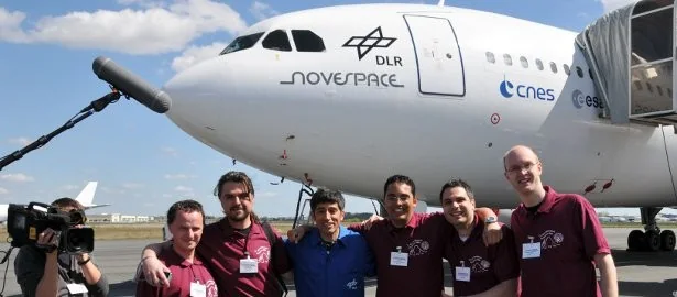
The 13th DLR Parabolic Flight Campaign in 2008 included an electrical impedance tomography experiment associated with the Göttingen research team [4]. The Airbus A300 ZERO-G provided repeated transitions between approximately 1 g, increased apparent gravity during pull-up/pull-out, and near-weightlessness during the ballistic part of the trajectory. The exact acceleration trace of this particular experiment is not reproduced here.

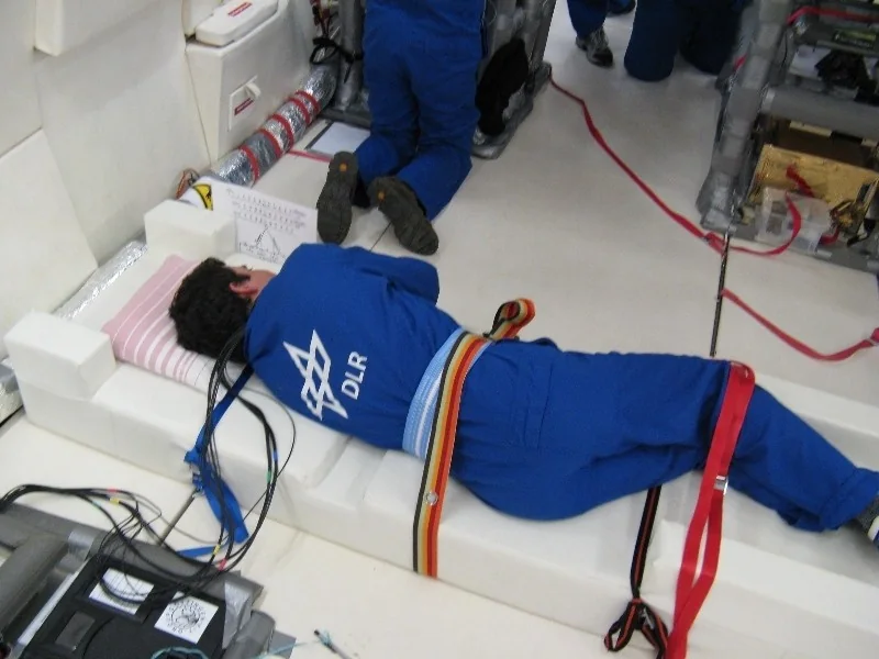
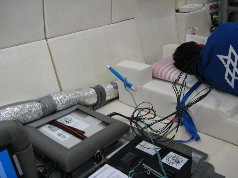
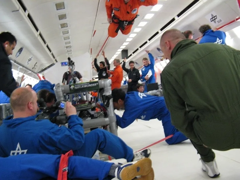
During the experiment, the subject must remain safely secured while electrode measurements are acquired. Cables, electrodes and associated equipment must also be protected against movement during the gravity transitions. Data analysis then has to distinguish physiologically meaningful impedance changes from artifacts caused by motion, electrode contact or changes in posture.
WHAT CHANGING GRAVITY CAN REVEAL
On Earth, gravity contributes to regional differences in lung mechanics, perfusion and the distribution of gas. The direction and magnitude of these effects depend on posture and breathing conditions. A parabolic flight offers a transient perturbation: researchers can compare EIT-derived ventilation or aeration indices across the different apparent-gravity phases.
Earlier published studies from the Göttingen group demonstrated that EIT can detect gravity-dependent changes in regional lung ventilation [1]. Subsequent work investigated absolute EIT and regional aeration under weightlessness and hypergravity [3]. Those publications provide the scientific context for this article; their published results must not be interpreted as measurements extracted from the photographs of the 2008 campaign.
INSIDE THE AIRCRAFT — EXPERIMENTAL PHOTOGRAPHS
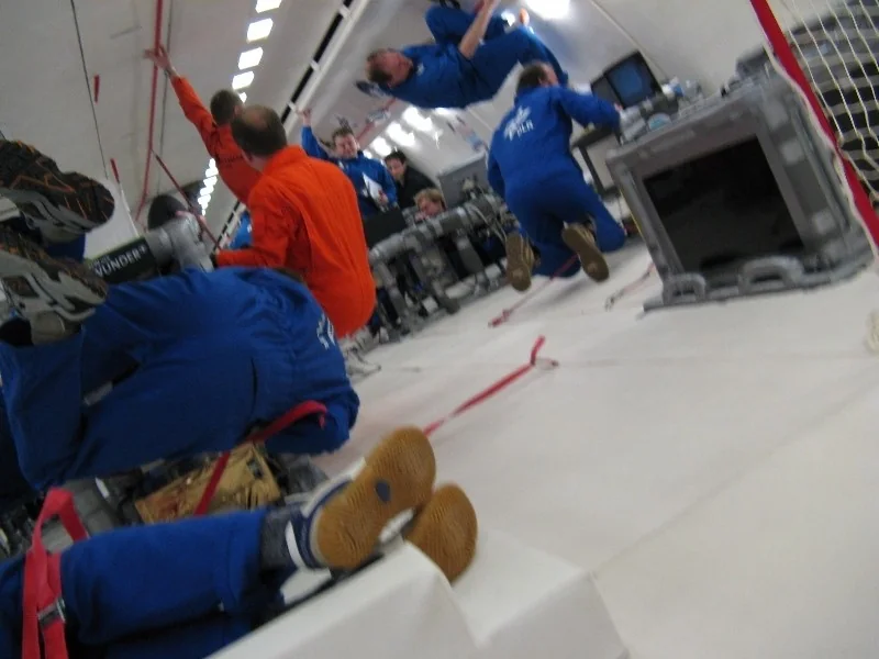
The following photographs document the experimental setting and the unusual working conditions of a parabolic-flight campaign. They are documentary images, not reconstructed EIT datasets.

CONCLUSION
Electrical impedance tomography connects elementary electrodynamics with an inverse problem and a demanding physiological experiment. Parabolic flight adds a controlled, short-duration change in apparent gravity. Together, they provide a way to investigate regional lung behavior under conditions that cannot be maintained in an ordinary laboratory. The central distinction remains: the electrodes measure electrical boundary signals; lung images are model-based reconstructions of those signals.
REFERENCES
[1] Frerichs, I., Dudykevych, T., Hinz, J., Bodenstein, M., Hahn, G. & Hellige, G. (2001). Gravity effects on regional lung ventilation determined by functional EIT during parabolic flights. Journal of Applied Physiology. DOI: 10.1152/jappl.2001.91.1.39.
[2] Hahn, G., Dittmar, J., Just, A. & Hellige, G. (2008). Improvements in the image quality of ventilatory tomograms by electrical impedance tomography. Physiological Measurement, 29(6), S51–S61. DOI: 10.1088/0967-3334/29/6/S05.
[3] Hahn, G., Just, A., Hellige, G., Dittmar, J. & Quintel, M. (2013). How absolute EIT reflects the dependence of unilateral lung aeration on hyper-gravity and weightlessness? Physiological Measurement. PubMed record.
[4] Iwanowski, I. (2008). 13th Parabolic Flight — Electrical Impedance Tomography. Project archive. Project documentation.
Image credits: Original parabolic-flight photographs from the participant archive; opening illustration AI-generated. Published EIT findings are cited independently and are not presented as newly analyzed experimental data.